166,000 Neurons, Fully Mapped: The Complete Fruit Fly Connectome Lands in Cell
Eighteen years after Janelia bet it could map a complex brain, researchers with Google and Cambridge publish the full male fruit fly connectome — 166,000 neurons and ~125 million synapses — the largest brain map ever assembled.
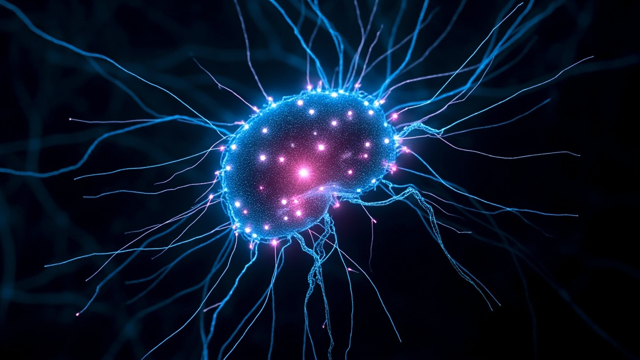
On September 3, 2026, a collaboration led by HHMI’s Janelia Research Campus, together with the MRC Laboratory of Molecular Biology, the University of Cambridge, and Google Research, published in Cell the complete connectome of an adult male fruit fly’s central nervous system. The wiring diagram covers more than 166,000 neurons and roughly 125 million synapses spanning the brain, both optic lobes, and the ventral nerve cord — making it the largest brain map by neuron count ever completed.
It is the culmination of an 18-year effort that many scientists once considered quixotic, and it hands the neuroscience community something it has never had before: a complete, searchable circuit diagram linking sensory perception all the way through to behavior, in an animal complex enough to navigate, court, fight, and learn.
Why a fruit fly brain matters
The fruit fly, Drosophila melanogaster, occupies a sweet spot in neuroscience. It performs sophisticated behaviors — goal-directed navigation, courtship songs, aggressive displays — with a nervous system small enough to map exhaustively. Before this week, the only complete animal connectomes were the 302-neuron worm C. elegans (finished in 1986 after over a decade of hand-drawn electron microscopy) and the adult female fly brain, completed by the FlyWire consortium in 2024 with roughly 139,000 neurons.
The new male map goes further than the female brain map in one crucial respect: it includes the ventral nerve cord — the fly’s spinal-cord analog — plus both optic lobes in their entirety. That means researchers can now trace an unbroken circuit from a photoreceptor in the eye, through the optic lobe, into the central brain, and out through the nerve cord to the motor neurons that actually move the legs and wings. Sensation to action, end to end.
A 1,000-fold efficiency bet that paid off
When Janelia’s founding executive director Gerry Rubin proposed mapping the fly brain in 2008, the skeptics had math on their side. The team’s own estimate was that the project would take 500 people working for 10 years with the technology of the day. The C. elegans map had taken more than a decade for a brain three orders of magnitude smaller.
What made it feasible was a relentless push on two fronts. Janelia’s Harald Hess and colleagues spent years optimizing and scaling electron microscopy to image neuronal tissue at nanometer resolution. Meanwhile, collaborations with Google Research produced computational reconstruction methods — the flood-filling neural networks that automatically segment each neuron from terabytes of imaging data — that eventually drove a more-than-1,000-fold increase in connectome generation efficiency.
The intermediate milestone came in 2020 with the “hemibrain,” a 25,000-neuron map of half a fly brain that was, at the time, the largest and most detailed connectome ever built. It proved the concept and triggered a wave of imitation: research teams worldwide now pursue wiring diagrams of increasingly complex brains. “There’s a point where people lack imagination, but they finally see something and say, ‘I got it,’” Rubin told HHMI. “That’s what the hemibrain did for neuroscientists.”
What the map is already revealing
Because a complete female fly brain connectome already exists from 2024, the publication unlocks something genuinely novel: the first side-by-side comparison of complete male and female brains of the same species. Early findings from that comparison are striking:
- Shared machinery, different routing. The core circuits for sensation and movement are largely common to both sexes. Sex-differentiated neurons cluster in higher brain areas rather than being scattered throughout.
- Circuit “switches.” Incoming sensory signals get rerouted into different behavioral pathways depending on sex — a likely mechanism for how the same stimulus (say, a pheromone cue) triggers courtship in males but a different response in females.
- A male-specific command network. Roughly 100 male-specific interneuron cell types form a highly intertwined network in the central brain that appears to coordinate most male-specific behaviors — mating and aggression among them.
Beyond sex differences, Janelia researchers say the map is already yielding new insights into the fly’s visual and taste systems, and into how the insect carries out goal-directed navigation.
The AI connection
The relationship between connectomics and AI runs in both directions. Google Research’s machine-learning pipelines were essential for reconstructing neurons at a scale no human team could trace by hand — a case study in AI accelerating fundamental biology. In return, complete connectomes are one of the only existing examples of a “neural network” whose architecture, learned over hundreds of millions of years of evolution, is fully specified. Neuroscientists and AI researchers alike study them for clues about wiring efficiency, recurrent motifs, and how compact circuits (a fly’s entire brain would fit in a fraction of a modern language model’s parameter count) generate such rich behavior.
HHMI is explicit about the AI angle in its next phase: an AI@HHMI project is now building “molecularly annotated” connectomes that record not just which neurons connect, but what chemical signals they exchange, and another project led by Janelia’s Misha Ahrens is combining a larval zebrafish connectome with whole-brain activity recordings to build predictive models of brain function.
What comes next
Janelia is not stopping at the fly. The campus is completing a whole-brain connectome of the larval zebrafish — a vertebrate — that will include neuronal activity data recorded while the fish behaves, and has begun work on the adult Danionella, a fish that stays transparent its entire life, making it ideal for pairing wiring diagrams with live imaging. The stated long-term goal is a mechanistic account of how a vertebrate brain generates complex behavior — information that could ultimately illuminate how disorders like Alzheimer’s, autism, and depression arise from miswired circuits.
The datasets are already public, downloadable and explorable through Google’s Neuroglancer tooling, which means the real payoff — thousands of discoveries made by scientists who never touched the underlying microscopes — starts now.
The bigger picture
The completion of the fly connectome is a reminder of how computing has changed the texture of biology. An 18-year project that began with hand-tuned microscopes ends with neural networks segmenting synapses, public cloud-hosted datasets, and a field — connectomics — that barely existed two decades ago now producing complete nervous systems as reference material. The worm gave neuroscience its first wiring diagram. The fly gives it its first full behavioral control system. The fish, and eventually the mouse, are next.
For a field often accused of chasing benchmarks, this is the other kind of AI story: machine learning quietly enabling a landmark of basic science. 166,000 neurons down, 86 billion to go.
Sources
- [1] https://www.hhmi.org/news/scientists-complete-full-map-fruit-fly-brain-connectome
- [2] https://research.google/blog/a-connectomics-milestone-mapping-the-complete-male-fruit-fly-brain/
- [3] https://www.eurekalert.org/news-releases/1141929
- [4] https://www.cam.ac.uk/research/news/comparison-of-male-and-female-fly-brains-is-unlocking-the-secret-workings-of-the-mind
- [5] https://www.janelia.org/news/researchers-reveal-connectome-of-the-male-fruit-fly-central-nervous-system