A Mouse Cortex Made of Human Cells: Stanford's Xenocortical Mice Redefine Brain Research
Stanford scientists bioengineered mice missing most of their cerebral cortex, then transplanted human cortical organoids that grew to fill over 90% of the empty cortex volume, connected to the spinal cord, and even produced rare von Economo neurons never before seen in a lab.
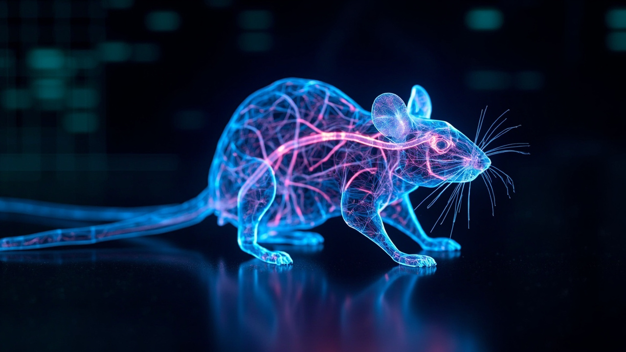
On September 16, 2026, a team at Stanford Medicine published a study in Nature that reads like science fiction executed with a pipette: mice whose cerebral cortex — the outer “rind” of the brain responsible for cognition, language, attention, and decision-making — is composed overwhelmingly of living human neurons. The researchers call them “xenocortical mice.” Three months after surgery, more than 90% of the cortical tissue in the animals’ brains was human.
The study, led by Sergiu Pasca — the Bonnie Uytengsu and Family Director of the Stanford Brain Organogenesis Program — is the most dramatic demonstration yet of organoid transplantation technology, and it opens a working laboratory model for conditions that have resisted study for decades: schizophrenia, epilepsy, profound autism, and cerebral palsy.
The problem: human brains resist study
The stubborn fact behind this work is that living human brain tissue is nearly always inaccessible. One in 20 American adults lives with a severe psychiatric illness. More than 1 in 100 has schizophrenia, believed to result largely from brain-circuit abnormalities that predate birth. Roughly 1% of people remain epileptic throughout adulthood. One in every 218 American children meets the criteria for profound autism — a state requiring round-the-clock supervision, with measured IQs below 50, elevated vulnerability to epilepsy and self-injury.
Animal models have helped, but as Pasca puts it, “some biological features seem to be uniquely human.” A decade of work on cortical organoids — three-dimensional, self-organizing clusters of human brain tissue grown from reprogrammed skin cells — got researchers partway there. In 2022, Pasca’s team transplanted such organoids into newborn rats and used the model to pinpoint the molecular defect of Timothy syndrome and find a candidate drug, now heading toward phase 1/2 safety trials.
But there was a structural limit: the human graft and the developing rat cortex were competing for the same turf, and host neurons matured faster than the human ones.
The leap: empty the cortex first
The new study’s central innovation flips the strategy. Instead of crowding human tissue into an intact rodent brain, the team first created a laboratory mouse strain whose own cortex largely never forms.
The genetic engineering is intricate. In cells expressing the pallial marker Emx1 on an immunocompromised SCID background, the researchers deleted Esco2, a sister chromatid cohesion gene. The “starter cells” that would normally give rise to the neocortex and certain other structures never get formed. The result: “apallial” mice that survive into adulthood in healthy condition despite missing most of their cortex and hippocampus, retaining a mere 2% of ordinary mice’s cortical content.
The empty space changed everything. The enlarged, fluid-filled cavities in newborn apallial mice proved a hospitable environment for human cortical organoids — each containing roughly 100,000 cells derived from healthy consenting donors, typically more than one graft per animal, surgically placed at two days of age.
The human tissue survived, thrived, and grew. Three months later, the grafts had expanded to fill more than 90% of the cortical volume. Human and mouse neurons were integrating into shared circuits. Human cells projected into the superior colliculus, sparsely into the spinal cord, and showed organized calcium and electrophysiological activity. Locomotion remained broadly intact, with only selective gait and spontaneous-behavior differences. Three to six months after transplant, the xenocortical mice performed generally like same-age normal peers on behavioral tests.
The cell nobody could grow
Then came the discovery nobody expected. Inside the human tissue, the researchers found detectable numbers of von Economo neurons — VENs — the large, cigar-shaped nerve cells situated in humans’ fronto-insular and anterior cingulate cortex, regions tied to social awareness and decision-making.
VENs had previously been observed only in postmortem brain tissue. They had never been generated in culture and had never turned up in earlier, more crowded transplant experiments. They are extraordinarily rare — roughly one in every 90,000 cortical neurons — and are known from other large-brained, highly social species: great apes, elephants, dolphins, whales.
Their sudden appearance matters because they are particularly vulnerable in frontotemporal dementia, a neurodegenerative disease that can begin in midlife. In some forms, VENs are markedly reduced, and early symptoms — changes in social behavior, personality, language — often emerge before prominent memory problems. “Now we can generate these rare cells from a healthy person and study them in a living, behaving animal,” Pasca said. “Or we can derive cells from patients with frontotemporal dementia, transfer them into these mice, test the animals for cellular or circuit-level correlates of behavioral disability, and then screen therapeutic candidates.”
A disease model that behaves
The study includes a proof-of-concept with direct clinical stakes. Xenocortical mice exposed to five hours of low oxygen suffered substantial damage to their human cortical tissue and subsequently showed trouble sustaining a steady gait and maintaining balance — reminiscent of children with cerebral palsy. Normal and apallial mice were virtually unaffected by the same exposure.
That differential vulnerability is the point. Oxygen deprivation during pregnancy or around birth is a known cause of cerebral palsy, which affects 3 in 1,000 Americans and is a risk factor for epilepsy and autism. For the first time, the specific susceptibility of human neurons to hypoxia can be observed in a living, behaving system — and, eventually, attacked with candidate therapies.
The ethics ledger
A result this provocative did not proceed without scrutiny. Pasca’s team spent years gathering input from ethicists, primate and human cortical biologists, patient advocates, philosophers, and legal scholars. In November 2025, Pasca organized a conference at Asilomar, California — the site of the famous 1975 recombinant-DNA gathering — to debate the ethical implications of human stem cell models and transplantation.
His framing of the moral calculus is pointed: “An overriding argument questioned the ethics of not conducting this research in the face of the suffering of hundreds of millions of people afflicted with neurological disorders that today are uncurable but tomorrow could yield treatments we discover by using this model.”
Commentators have already begun drawing lines. Neuroscientist Anil Seth called the work a synthetic-biology milestone while raising unresolved consciousness questions; philosopher Jonathan Birch separately flagged concern that such research creates hosts with unknown welfare needs rather than reducing animal research overall.
Why it matters
The xenocortical mouse is not a mouse with a human mind, and the researchers are careful not to claim one. It is something more useful: a chimeric platform where uniquely human neural biology — cortical cell diversity, long-range projections, VENs, hypoxia vulnerability — develops inside an intact nervous system that can be observed, measured, and experimentally manipulated.
Stanford’s Office of Technology Licensing holds patents for generating cortical organoids, with Pasca listed as an inventor, and a provisional patent application for the transplantation method itself. The study was funded by the Wu Tsai Neuroscience Institute, the Kwan Funds, the Senkut Funds, and the Brain & Behavior Research Foundation.
For the AI-adjacent world watching from the outside, the study is also a reminder that “understanding intelligence” is not only a computational project. The most complete maps of human neural development are still being drawn not in silicon but in the brains of mice — rebuilt, cell by cell, from our own.
Sources
- [1] https://www.nature.com/articles/s41586-026-11032-2
- [2] https://med.stanford.edu/news/all-news/2026/09/model-to-study-brain-development.html
- [3] https://www.technologyreview.com/2026/09/16/1144210/meet-a-mouse-whose-brain-cortex-is-made-up-of-human-cells/
- [4] https://www.thetransmitter.org/spectrum/human-brain-organoids-flourish-in-emptied-mouse-cortex/
- [5] https://www.sciencealert.com/scientists-grew-human-brain-tissue-inside-mice-heres-what-happened
- [6] https://gizmodo.com/scientists-grew-more-human-brain-tissue-in-a-mouse-than-ever-before-and-people-have-questions-2000812503